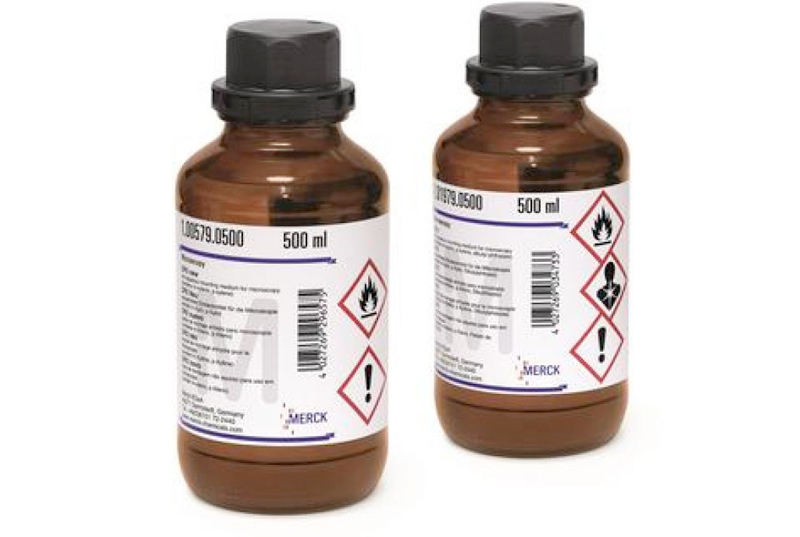

محلول گیمسا (محلول آزور، ائوزین، متیلن بلو در متانول)
• برای رنگ آمیزی لام های خونی، نمونه های مغز استخوان، بیوپسی معده، برش های بافت پارافینی و ...
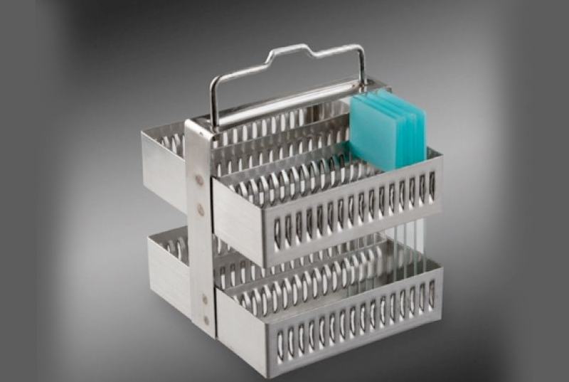
• بطری شیشه ای 250 و 1000 میلی لیتری
• محلول آماده برای مصرف
• محلول آزور، ائوزین، متیلن بلو
• دارای الکل متانول
محلول رنگ آمیزی گیمسا (محلول آزور، ائوزین، متیلن بلو در متانول)
محلول رنگ گیمسا یک محلول کلاسیک جهت رنگ آمیزی لام های خونی و نمونه های مغز استخوان و برش های بافت پارافینی می باشد. گلبول های قرمز رنگ صورتی، پلاکت رنگ صورتی کمرنگ، سیتوپلاسم لنفوسیت رنگ آبی آسمانی، سیتوپلاسم مونوسیت رنگ آبی کمرنگ و هسته لکوسیت رنگ ارغوانی به خود می گیرند. همچنین از محلول گیمسا برای رنگ آمیزی قارچ ها و باکتری کلامیدیا و همچنین رنگ آمیزی سلول های Mast و کروموزوم ها نیز استفاده می شود.
Giemsa Stain Solution (Solution of eosin, methylene blue and azure dyes in methanol)
Giemsa stain is a member of the Romanowsky group of stains and is a classic differential stain for peripheral blood smears and bone marrow specimens. Giemsa can also be used to stain bacteria (such as H.pylori), malaria and other parasites. Giemsa stain used for the preparation of cyto-histological samples for optical microscopy.
Giemsa's staining is one of the standard procedures in histology, hematology, cytology and bacteriology. The Giemsa's azur-eosin-methylene blue dye is a dry dye that is used for the preparation of a staining solution. This solution can be used for the staining of blood and bone marrow smears, paraffin sections, spirochaets and clinical-cytological specimens of human origin. The stain demonstrates various cells with their morphological features better than e.g. with H&E stain in histology and it can also be used to detect Helicobacter pylori in gastric tissue biopsies. Alternatively, for some applications, among others, May-Grünwald's eosin-methylene blue can be used as a substitute for Giemsa's solution.
Application
Reagent used with May Grünwald for the staining of different kind of cells in blood and bone marrow smears. This staining is also used to highlight the Helicobacter pylori in histology.
For the execution of the staining method is required the use of May Grünwald solution.
Principle
Two dyes are used one after the other:
- May Grünwald solution, consisting of eosin-methylene blue, stains nuclei blue and basophil cytoplasm in pinkish red;
- Giemsa solution, complex consisting of methylene blue chloride, eosin-methylene blue and azure II eosinate, improves the intensity of nuclear staining and the capacity to show selectively cellular structures.
Method
To appreciate results always remember two factors: pH of washing waters and dilution buffer have a strong influence on final colour chart; intensity of stain may vary according to differentiation time.
1) Air dry smears
2) May Gruenwald solution 5 minutes
3) Wash in tap water 1 minutes
4) Giemsa working solution 15 minutes
5) Wash in tap water 1-2 minutes
6) Air dry
*Preparation of Giemsa working solution:
Dilute Giemsa solution in distilled water at ratio 1 : 10 (1 part of Giemsa solution + 9 parts of distilled water).
Results
Nuclei....................................................................violet red purple
Lymphocyte cytoplasm........................................different blue tonalities
Monocyte cytoplasm............................................blue – grey
Eosinophil granulocytes (acidophil granules).....brick red – orange
Basophil granulocytes (basophil granules).........dark violet
Neutrophil granulocytes (neutrophil granules)....pink – brown
Erythrocytes............................................................pink – grey