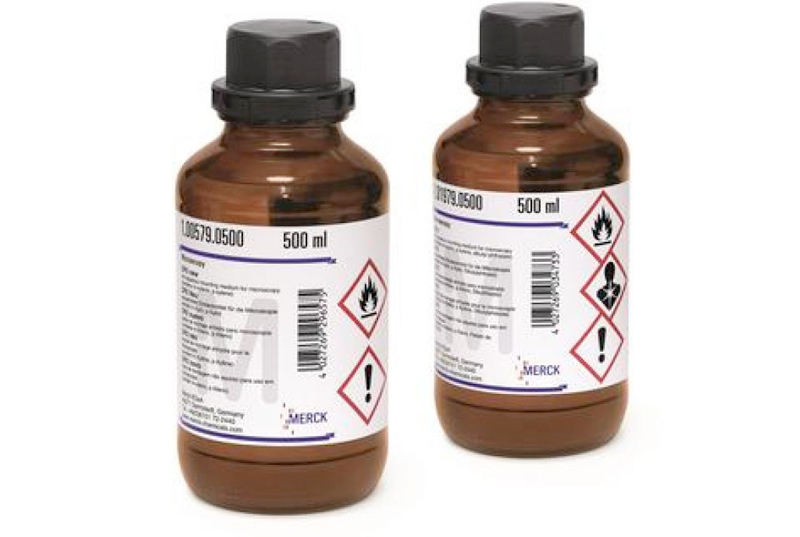
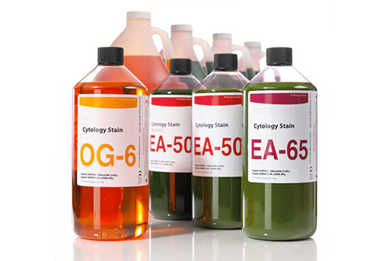
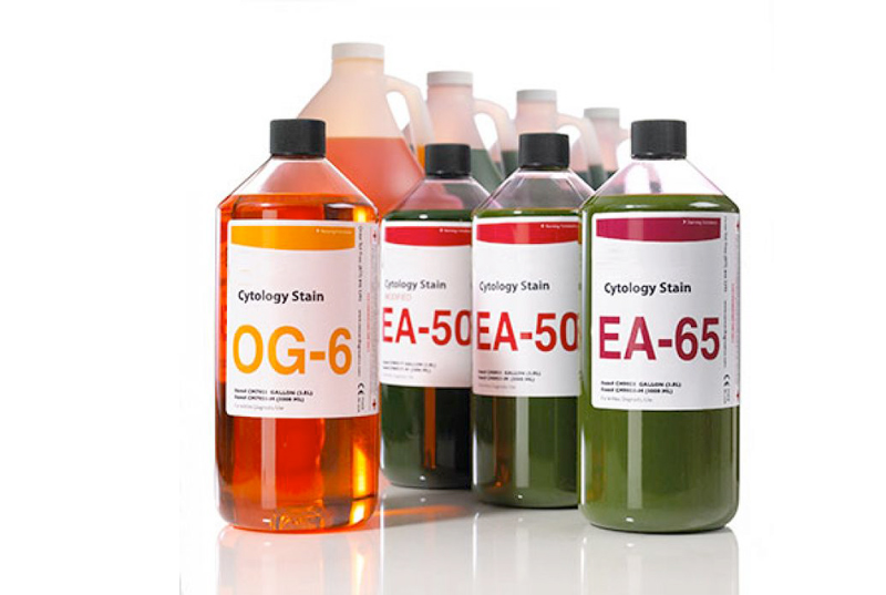
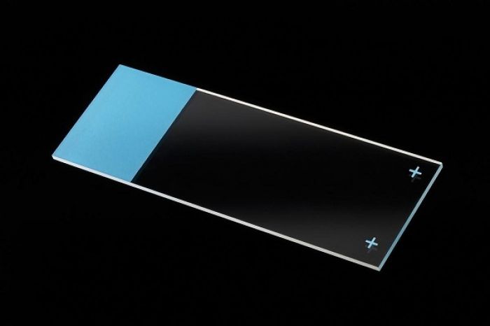
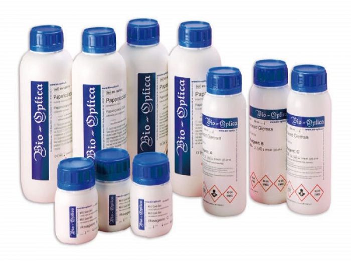
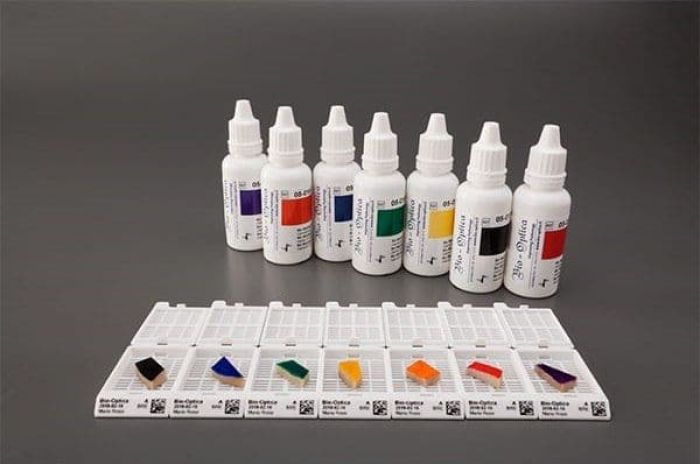
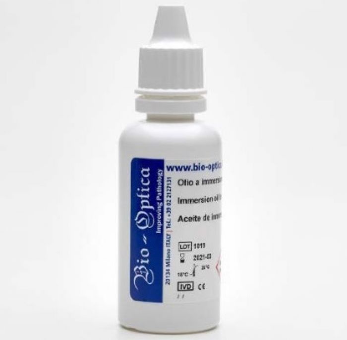
محلول رایت (محلول ائوزین، متیلن بلو در متانول)
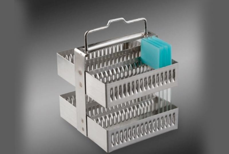
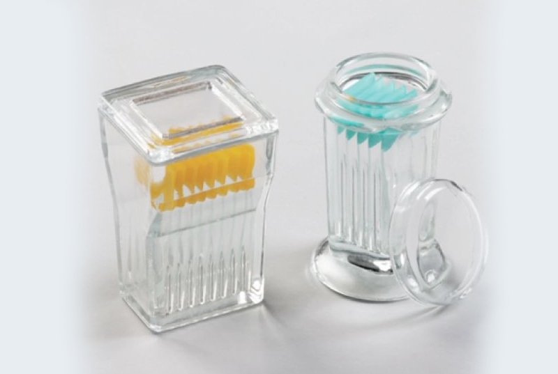
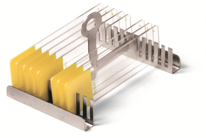
• برای رنگ آمیزی لام های خونی، نمونه های مغز استخوان، بیوپسی معده، برش های بافت پارافینی و ...
• بطری شیشه ای 250 و 1000 میلی لیتری
• محلول آماده برای مصرف
• محلول ائوزین، متیلن بلو
محلول رنگ آمیزی رایت (محلول ائوزین، متیلن بلو)
محلول رنگ رایت یک محلول کلاسیک جهت رنگ آمیزی لام های خونی و نمونه های مغز استخوان و برش های بافت پارافینی می باشد. گلبول های قرمز رنگ صورتی، پلاکت رنگ صورتی کمرنگ، سیتوپلاسم لنفوسیت رنگ آبی آسمانی، سیتوپلاسم مونوسیت رنگ آبی کمرنگ و هسته لکوسیت رنگ ارغوانی به خود می گیرند. همچنین از محلول گیمسا برای رنگ آمیزی قارچ ها و باکتری کلامیدیا و همچنین رنگ آمیزی سلول های Mast و کروموزوم ها نیز استفاده می شود.
نحوه استفاده از محلول رایت
1- گستره را به وسیله متانول فیکس نمایید.
2- روی سطح لام یک میلی لیتر رنگ بریزید.
3- بعد از 5 دقیقه یک میلی لیتر آب مقطر به سطح لام اضافه نموده به طوری که آب مقطر و رنگ مخلوط شوند.
4- بعد از گذشت 5 دقیقه لام را با آب شسته و بعد از خشک شدن زیر میکروسکوپ بررسی نمایید.
Wright's Solution
Wright's stain is a histologic stain that facilitates the differentiation of blood cell types. It is used primarily to stain peripheral blood smears and bone marrow aspirates which are examined under a light microscope. In cytogenetics it is used to stain chromosomes to facilitate diagnosis of syndromes and diseases.
Wright's stain is classically a mixture of eosin (red) and methylene blue dyes.
It is named for James Homer Wright, who devised the stain, a modification of the Romanowsky stain, in 1902. Because it distinguishes easily between blood cells, it became widely used for performing differential white blood cell counts, which are routinely ordered when conditions such as infection or leukemia are suspected.
The related stains are known as the buffered Wright stain, the Wright-Giemsa stain (a combination of Wright and Giemsa stains), and the buffered Wright-Giemsa stain, and specific instructions depend on the solutions being used, which may include eosin Y, azure B, and methylene blue (some commercial preparations combine solutions to simplify staining). The May–Grünwald stain, which produces a more intense coloration, also takes a longer time to perform.
Urine samples stained with Wright's stain will identify eosinophils, which can indicate interstitial nephritis or urinary tract infection.